

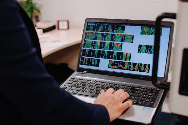
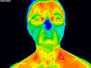
Thermography is a non-invasive, diagnostic tool that takes digital images of the body which are sent to a board of Thermography trained Doctors to analyse patterns and changes in the skin’s surface temperature.
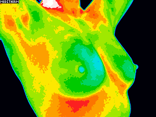
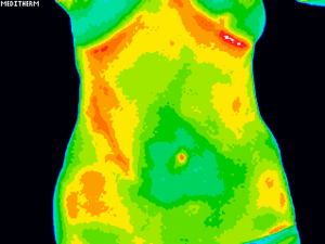
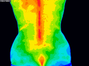
There is now a consensus that chronic inflammation is a clinical marker for a compromised immune system and the onset of chronic disease – even as the initial inflammatory condition may be asymptomatic for an extended period of time. Areas of inflammation present with higher temperatures and are clearly identified from the thermal maps, or thermograms, generated by thermography imaging.
Clinical Thermography is a simple test of physiology that relies on the sympathetic nerve control of skin blood flow and the ability of the sympathetic system to respond and act to pathology anywhere in the body.
This test of function relies on the sympathetic nerve control of skin blood flow and the ability of the sympathetic system to respond and react to pathology anywhere in the body. In simple terms, this test can indicate where there are inflammatory conditions in the body and show up early signs of abnormal cell development.
What can Thermography see?
What do I do with my Thermography report and images?
It is important that you carefully read the report and look up any unfamiliar medical terms. If ‘clinical correlation’ is suggested, you need to take a proactive approach and follow up on this. It could be a visit to your GP and/or another healthcare provider such as a physiotherapist, dentist, nutritionalist, naturopath or just taking a close look at your diet and lifestyle choices and initiating changes.
We promote a model of ‘self responsibility’ when it comes to health and wellness. If you need clarification on any of the findings, you can email us. The thermal findings from your report and images will be very useful for you and/or your chosen healthcare provider to help diagnose and monitor pain and/or pathology in any part of your body and provide useful information in planning a treatment program.
Can Thermography diagnose cancer?
Who interprets the Thermography images?
Should I use Thermography instead of a mammogram?
Thermography is an adjunctive test and does not replace mammography as each test provides different results and information. A mammogram will show structural abnormalities such as densities, calcifications and tumours. Whereas Thermography doesn’t show any structure but does show functional abnormalities like inflammation and vascular and lymph dysfunction. By using both modalities the highest rate of detection is going to be gained.
What conditions can be detected?
Is it Dangerous?
Thermography is a non-invasive procedure where thermal images are taken of the body with a Thermal Image Camera.
Can I have a study if I am breastfeeding?
Due to the amount of thermal activity surrounding the breasts during breastfeeding it is not recommended to have an image taken during this time as the breastfeeding process will mask any potential abnormalities. It will take three months after finishing breastfeeding to have an accurate thermal image taken.
The same applies to pregnancy. It is best to wait at least 3 months after giving birth before booking in for your Thermography study.
Is Thermography covered by private health insurance?
What are the benefits of Thermography?
This can be used by individuals to take a preventative and proactive approach to their health and well-being. Thermography can highlight areas that could warrant further investigation long before symptoms may be felt.
As there are no harmful rays involved in the imaging process, individuals can safely and regularly use Thermography to monitor the efficacy of an ongoing treatment program.
Why do I need a follow up breast study?
The reason for the follow up breast study is to establish your accurate and unique stable thermal baseline which will be used for future comparative studies. This cannot be done with just one study as we would have no way of knowing if this is your normal pattern or if it is actually changing at the time of your first study. By comparing the two studies 90 days apart the interpreting Doctors would be able to judge if your breast physiology is stable and suitable to be used as your normal stable baseline. There needs to be a minimum 3 month gap as this is the period of time it takes for blood vessels to show any change or not.
There is a very useful case study which can be viewed on the Island Images Gallery of images that demonstrates the need for the 3 month follow up. If you don’t have your follow up, your study is not complete. This is only done once, annual or bi-annual studies only require one appointment.
Payment plans available for non-discounted Thermography sessions.